Research Focus:
Cancer covers a broad spectrum of diseases, in every tissue of the body. Tissues are composed of cells, which normally grow slowly, under the tight control of a network of regulatory genes.
The slow accumulation of activating mutations in growth genes, and inactivating mutations in suppressor genes, eventually allows a cell to grow out of control. Relapse is due to the development of resistant cells, rather than the escape of sensitive cells, suggesting the need for new approaches to treatment of the disease.
This laboratory is developing sequence-specific oligonucleotides against cancer genes and neurological genes for use as diagnostics and therapeutics. The cancer gene mRNAs being studied include the CCND1, HER2, IGF1R, KRAS2, and MYCC in breast cancer, prostate cancer, colon cancer, lung cancer, and brain cancer.
In a new direction, we have begun to knock down two microRNAs, miR-17 and miR-21, which are overexpressed in triple negative breast cancer cells. Micro RNA precursor duplexes were thought to 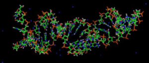 include an active guide strand and an inactive passenger strand.
However, we discovered passenger strand activity in triple negative breast cancer cells, when anti-miR-17-5p depressed PTEN and PDCD4 protein, instead of raising them. We have observed that nuclease-resistant sequences that specifically block miR-17 or miR-21 interdict triple negative breast cancer cell growth.

To translate microRNA blockade into clinical cancer management, Thomas Jefferson University has licensed our technology to Bound Therapeutics LLC.
To move our approaches into the clinic, we must identify the most efficacious antisense target sequences, their mechanisms and physiological effects. We must design and synthesize potent RNA analogs capable of surviving in the bloodstream following administration, and we must determine their structures bound to cellular organelles.

